

Recurrent pregnancy loss (RPL), clinically recognized as recurrent miscarriage, remains one of the most agonizing challenges in modern reproductive medicine. For American couples striving to build a family, facing two or more consecutive pregnancy losses before the 20th week of gestation introduces deep emotional trauma and complex clinical questions. While chromosomal abnormalities, thrombophilias, and endocrine disorders account for a significant portion of losses, anatomical uterine pathology represents a primary underlying factor in up to 25% of cases evaluated by reproductive endocrinologists.
The uterine cavity serves as the physiological foundation for blastocyst implantation, placentation, and embryonic development. Any structural disruption within the endometrium or myometrium can compromise uterine receptivity, severely disrupting early vascularization. Understanding the specific recurrent pregnancy loss causes originating from structural anomalies is vital to transitioning from recurrent grief to effective clinical intervention.
Categorizing uterine factors in repeated miscarriages
Uterine causes of repeated miscarriages are broadly classified into congenital Müllerian anomalies (present from birth) and acquired structural uterine pathology (developed later in life).
Congenital Müllerian Anomalies
Müllerian duct anomalies occur during embryogenesis when the female reproductive tract fails to fuse or reabsorb properly.
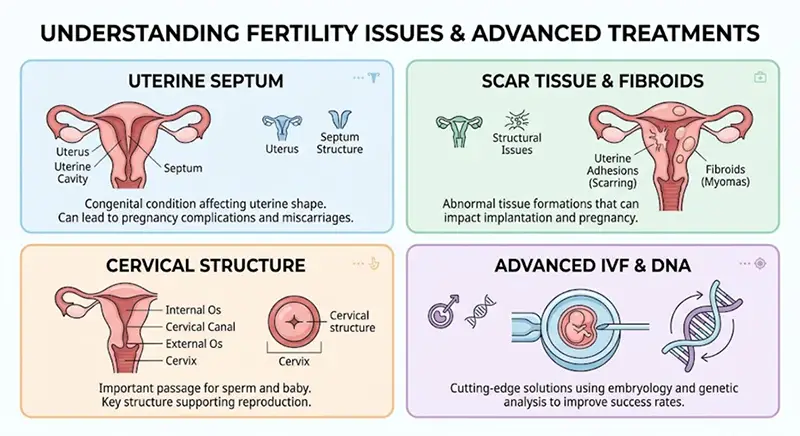
- Septate Uterus: The most prevalent congenital anomaly associated with recurrent spontaneous abortion. A fibrous, poorly vascularized septum divides the uterine cavity. When an embryo implants directly on the septum, inadequate blood supply frequently causes early embryonic demise.
- Bicornuate and Didelphic Uterus: Resulting from incomplete fusion of the Müllerian ducts. These anomalies create structural limitations and reduced endometrial surface area, leading to mid-trimester miscarriages or severe preterm labor.
- Unicornuate Uterus: Represents a single-horned uterus with reduced vascular capacity, significantly elevating the risk of early pregnancy loss.
Acquired uterine lesions
Acquired uterine conditions alter the endometrial environment through trauma, infection, or abnormal tissue growth:
- Asherman Syndrome (Intrauterine Adhesions): Characterized by bands of scar tissue crossing the uterine cavity, often resulting from previous dilation and curettage (D&C), postpartum infection, or uterine surgery. Severe intrauterine synechiae obliterate the functional endometrial layer, preventing healthy implantation.
- Submucosal Leiomyomas (Fibroids): Myomas that distort the endometrial cavity physically obstruct blastocyst attachment and induce chronic localized inflammation and abnormal uterine contractions.
- Chronic Endometritis: A persistent, low-grade inflammatory condition of the endometrial lining caused by bacterial pathogens. It alters mucosal immune tolerance, triggering early gestational failure.
Cervical insufficiency and vascular receptivity factors
Cervical insufficiency (incompetence) involves painless cervical dilation during the second trimester, resulting in spontaneous membrane rupture and pregnancy loss. Diagnostic guidelines and evidence-based standards established by the American Society for Reproductive Medicine (ASRM) and scientific peer-reviewed studies indexed on NCBI PubMed emphasize evaluating cervical length via serial transvaginal ultrasound.
Furthermore, Doppler ultrasound assessment of uterine artery blood flow reveals that elevated vascular resistance can impair endometrial thickness, starving the developing placenta of oxygen and metabolic support.
Advanced diagnostic testing protocols
Accurately evaluating the uterine cavity requires state-of-the-art diagnostic modalities:
- Diagnostic Hysteroscopic Evaluation: Considered the clinical gold standard. It allows direct visualization of the endometrial cavity, precise grading of intra-cavity adhesions, and immediate surgical correction of septa or polyps.
- 3D Transvaginal Ultrasonography: Provides high-resolution volumetric rendering of the outer uterine contour and inner cavity, effortlessly distinguishing between septate and bicornuate uteri.
- Saline Infusion Sonohysterography (SIS): Uses sterile saline solution to distend the endometrial cavity during ultrasound, highlighting subtle mucosal defects and submucosal fibroids.
- Endometrial Biopsy & Immunohistochemistry (CD138): Essential for diagnosing chronic endometritis by identifying plasma cell infiltration in the stroma.
Medical interventions and reproductive surgeries
Addressing uterine factors of recurrent miscarriage relies on targeted medical and surgical therapies:
- Hysteroscopic Metroplasty: Surgical resection of a uterine septum restores normal cavity symmetry, dramatically increasing live birth rates in patients with septate uteri.
- Lysis of Intrauterine Adhesions: Careful hysteroscopic adhesiolysis combined with postoperative estrogen therapy helps regenerate healthy endometrial tissue in Asherman syndrome patients.
- In Vitro Fertilization (IVF) with Preimplantation Genetic Testing (PGT-A): Ensures that only genetically euploid embryos are transferred, eliminating chromosomal co-factors in couples with uterine structural challenges.
Overcoming irreversible uterine factors: gestational surrogacy
When uterine pathology is severe and non-reconstructible—such as refractory Stage IV Asherman syndrome, extensive myometrial scarring, or severe congenital uterine hypoplasia—carrying a pregnancy to term may be medically impossible. In these heartbreaking circumstances, gestational surrogacy provides a scientifically proven, safe pathway to biological parenthood.
For couples navigating complex fertility histories, exploring tailored programs for heterosexual couples offers comprehensive clinical management, gestational carrier matching, and complete legal protection.
Before initiating an international family-building journey, it is critical to understand the global legal landscape regarding where surrogacy laws exist to ensure parental rights are legally secured from birth.
Confronting recurrent pregnancy loss is an arduous journey, but with precise medical diagnostics, advanced surgical interventions, or gestational surrogacy, intended parents can realize their dream of welcoming a healthy child.